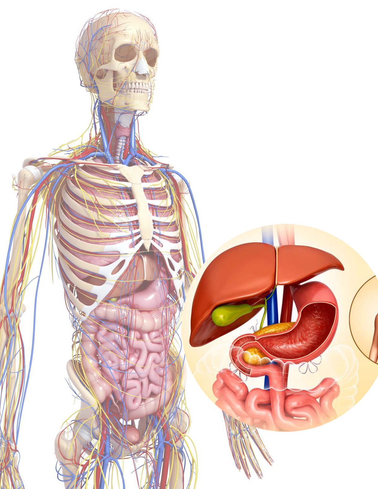
Digestive System
Lesson Plan
Grade 12

Elisabeth Ormandy, 2020.
Do not make copies and/or distribute the material contained in this document without
explicit, written permission.
2

TABLE OF CONTENTS
Curriculum alignment............................................................................. 4
Curriculum competencies & recommended tools........................... 5
Lesson plan overview............................................................................. 6
Lesson plan approach............................................................................. 7
Detailed lesson content & teaching notes......................................... 8
Digestive system major structures...................................................... 9
Food flow through the body................................................................ 11
Stomach anatomy................................................................................... 17
Pancreas anatomy.................................................................................. 19
Villi anatomy nutrient breakdown...................................................... 21
Interdependence of digestive and other organ systems............... 22
Hepatic portal system............................................................................ 24
The digestive system & homeostasis................................................. 25
Common digestive diseases................................................................. 27
Closing check-in & discussion.............................................................. 28
3
Elisabeth Ormandy, 2020.

4
Structure and function
Structural and functional interdependence
Maintenance of homeostasis
The following unit plan was created in accordance with the Canadian Council on
Animal Care’s recommendations to replace any present procedures involving the
use of animals in teaching, testing and research.
The Three Rs principle of Replacement states, if you can meet your scientific or
educational goals without the use of animals, it is your ethical obligation to use non-
animal methods. Grade 12 anatomy content is often taught using fetal pigs - here
we offer an effective and humane alternative.
This is in alignment with the public’s concern for animal welfare and a cultural
respect for animals passed down from the Aboriginal perspectives of the First
Peoples.
Elisabeth Ormandy created this unit plan and series of lesson plans for your use in
teaching life science content to Grades 12 based on the BC Science Curriculum.
These Humane Science Education materials were developed to provide equivalent
or greater standards in education for Canadian youth, without the use of animals.
Curriculum Alignment
This lesson plan can be used to create classes for Grades 12 based on the BC
Science Curriculum. Specific Big Ideas covered in this lesson plan include:
Grade 12 - Organ systems have complex interrelationships to maintain homeostasis.
ORGAN SYSTEMS:
We have recommended specific virtual anatomy tools to use to get the most out of
the lesson plan. You'll find links to those on pages 5&6.
Elisabeth Ormandy, 2020.

Lesson Plan Overview
Subject: Science
Unit Overview: Anatomy and Physiology
Unit Duration: ~90 minutes
Grade: 12
Big Idea: Organ systems have complex interrelationships to maintain homeostasis
Curricular Competencies
Analyze cause-and-effect relationships
Construct, analyze, and interpret graphs, models, and/or diagrams
Consider the changes in knowledge over time as tools and technologies have developed
Content
By the end of this lesson, students are expected to demonstrate understanding of
the following:
Digestive system:
Structure and function
Structural and functional interdependence
Maintenance of homeostasis
6 (or more) iPads or other tablets
6 (or more) 3D Anatomica workbooks
3D Anatomica:
https://3danatomica.com
3D4Medical Complete Anatomy: https://3d4medical.com
Hardware & Workbooks:
This inventory is for a regular in-person class - use x1 iPad/tablet per student for
responsible physical distancing. If teaching online, teachers can screen share their
iPad/tablet or desktop.
Recommended Software:
Recommended Education Tools
5
Elisabeth Ormandy, 2020.

Lesson Plan Overview
Topic: Organ systems have complex interrelationships to maintain homeostasis.
Homeostasis is maintained through physiological processes.
Content: The human digestive system: organs, structure and function
Goals
Objectives
Materials
Introduction
Development
Practice
Describe the function of the digestive system and its major organs.
Describe the relationships between the different components of the
digestive system.
Explain how the digestive system is interdependent with the
circulatory system.
Explain how the digestive system maintains homeostasis in the body.
Students will be able to:
After this lesson students will state the structure and function of each
organ/tissue in the digestive system and explain how the digestive
system is functionally interdependent with other body systems.
3DAnatomica
3D4Medical
Digestive System Workbook
Using the 3DAnatomica and/or 3D4Medical app(s), the teacher will
introduce the topic of cardiovascular organ structure and function.
What is the advantage of having specialized tissues in the digestive
system?
How does the digestive system help the body maintain internal
balance during exercise?
What are the impacts of external stimulants (e.g. alcohol, caffeine) on
the digestive system?
What lifestyle decisions would you make to improve your digestive
health?
How does the digestive system respond to infection by a pathogen?
Questions to support inquiry-based learning:
Students will work independently or in pairs to navigate 3DAnatomica
and/or 3D4Medical to learn about the structure and function of the
digestive organs.
6
Elisabeth Ormandy, 2020.

Split students into 6 groups.
Give each group a Digestive System workbook to refer to, and one (or more)
iPad(s) or tablet(s) with the 3D Anatomica app, and 3D4Medical Complete
Anatomy app loaded and ready to use.
Your introduction should include discussion of the function of the digestive
system, identifying its major components, and the vocabulary you would like
students to learn (~ 15 mins). Define homeostasis. Have the students follow along
using the 3D4Medical Complete Anatomy app.
Have students label "Major Structures" diagram on page 10 using 3D4 Medical.
Discuss sequence of organs and structures that food moves through within the
digestive system. Have students use the 3D Anatomica and 3D4Medical Complete
Anatomy app to explore the flow of food in their groups, filling their 3D Anatomica
workbook and/or handouts provided. This can be student or teacher led (40-45
mins).
Explore the "Structures in Detail" pages using the 3D4 Medical App. The students
can cut away at the structures in the app to locate structures that need to be
labeled.
Ask students to brainstorm ways the digestive system interacts with other
systems, and go over the specific examples provided
Discuss different ways the digestive system helps maintain homeostasis using
examples provided, then ask students to provide their own examples using what
they've learned.
Close the class with a 20-minute recap of what the students have learned, discuss
how the parts of the digestive system work together, and check for understanding.
Begin a conversation on ethics of animals in science using the questions provided.
Use x1 iPad for each student and proceed as per the directions above.
Lead the students through the digestive system by screen sharing your own
iPad/tablet or desktop with the 3D4Medical Complete Anatomy app installed,
filling out the tables, and labeling the models as you go.
Proceed as per the directions above.
If teaching regular in-person classes:
If teaching a physically-distanced class:
If teaching online:
Lesson Plan Approach
7
Elisabeth Ormandy, 2020.

Introduction to the Lesson
Include a First Nations land acknowledgement and ask students to reflect on what
respect for animals means to them. Provide an introduction to the apps and models
that will be used in class. Provide an overview of how to access 3D Anatomica
workbooks if teaching remotely.
What is Homeostasis? Discuss with Students
In biology, homeostasis refers to the body’s ability to maintain a stable internal
environment despite changes in external conditions.
Introduction to the Topic
Students will use the 3D4Medical Complete Anatomy app to explore the digestive
system at large. We recommend covering the function of the digestive system,
identifying the major components of the system, and discussing the vocabulary you
would like the students to learn early in the lesson.
Detailed Lesson Content & Teaching Notes
THE DIGESTIVE SYSTEM AT-A-GLANCE
8
Function
Components
Important vocabulary
The digestive system is responsible for taking whole foods and
turning them into nutrients and energy, which allows the body to
function, grow and repair itself.
Mouth, esophagus, stomach, small intestine, large intestine, rectum,
liver, pancreas, gallbladder.
Homeostasis, mouth, salivary glands, salivary amylase, palate,
esophagus, pharynx, epiglottis, esophageal sphincter, bolus,
peristalsis, stomach, chyme, mucus cells, parietal cells, peptic cells,
pepsin, HCl, peptides, pyloric sphincter, pancreas, mesenteric vessels,
exocrine, endocrine, pancreatic duct, alkaline, neutralize, basic,
amylase, peptidases, lipases, nucleases, glycogen, glucose, secretin,
gallbladder, bile, bile duct, small intestine, enzymatic hydrolysis,
duodenum, jejunum, ileum, villi, microvilli, cytoplasmic, amino acids,
lipoproteins, large intestine, cecum, iliac crest, appendix, ascending
colon, transverse colon, descending colon, rectum, feces, anus, liver.
Elisabeth Ormandy, 2020.

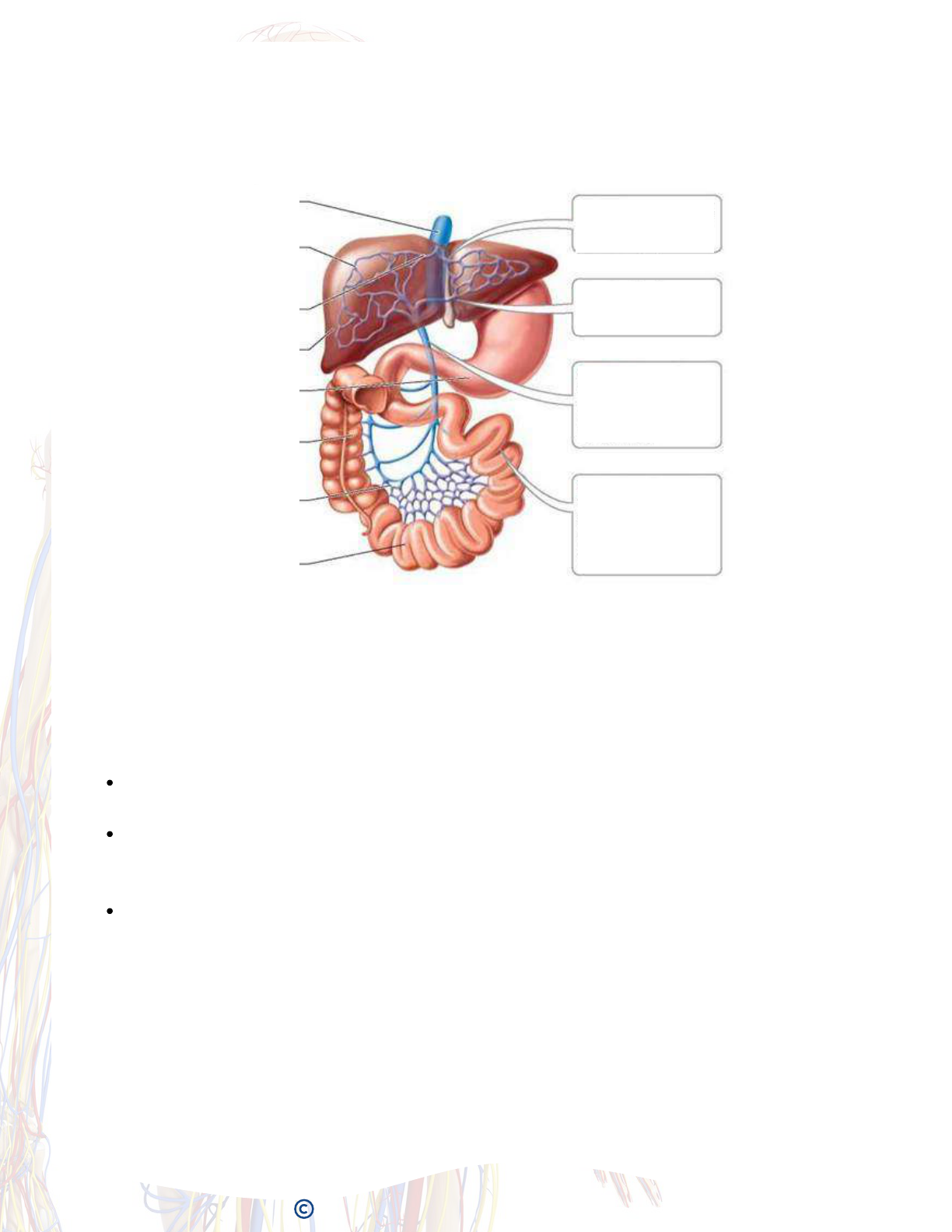
Digestive System Major Structures
(Teacher Copy)
9
Liver
Gallbladder
Pancreas
Duodenum
Esophagus
Stomach
Tranvserse
Colon
Jejunum
Descending
Colon
Rectum
Anus
Colon
Ileum
Ascending
Colon
Elisabeth Ormandy, 2020.

Digestive System Major Structures (Student Copy)
10
Elisabeth Ormandy, 2020.

Digestive System Food Flow (Teacher Copy)
Explore the path food moves through within the digestive system, noting the function
and structure of each major organ, using the apps provided, and fill out your workbook.
11
Order Organ Structure, function & information
1
2
3
Esophagus
Mouth
Stomach
Mechanical Digestion: chewing
Chemical Digestion: saliva breaks down carbohydrates/starches
2 Types of Digestion:
3 sets of salivary glands produce saliva to lubricate and break down the food.
Teeth: mix of canines and molars to masticate (tear/bite/grind/crush) the food into
smaller pieces.
Tongue: muscular organ that mixes the food with saliva and aids with swallowing.
Salivary Amylase: enzymes that digest the carbohydrates in the mouth.
Palate: forms the roof of the mouth.
From the mouth, food passes through the pharynx (5-6 inches long).
During swallowing, the sphincter muscles relax and raise the epiglottis, which
prevents the bolus from entering the trachea.
After bolus leaves pharynx, it enters the esophagus.
Peristalsis: contractions of the esophagus that move food bolus along in a wave
like motion.
Esophageal sphincter: separates esophagus from the stomach to prevent food
reflux.
Thick-walled, J-shaped organ, lies on left side of body, under diaphragm.
Stores food and mixes it with gastric juice.
Bolus enters the stomach, then is converted to semi-fluid, partially digested food called
chyme.
Stomach contents are extremely acidic (pH b/w 1.5-2.5).
Acidity breaks down food tissues, kills microorganisms & activates digestive enzymes.
3 layers of muscles churn and mix contents by contracting.
Pacemaker cells stimulate stomach contractions, which increase in number the fuller
the stomach.
Hunger pains= churning of an empty stomach.
Mucus lining of stomach contains inner gastric juice producing gastric glands.
Stomach empties into the first part of the small intestine (duodenum).
Pyloric sphincter at the bottom of the stomach controls this emptying.
Elisabeth Ormandy, 2020.

12
3.2
3.1
4
Pancreas
Gallbladder
Small
Intestine (SI)
Acinar Cells - secrete digestive juices which travel through pancreatic duct to
small intestine
Islets of Langerhans: secrete insulin and glucagon into blood
Location: just below the stomach
Two types of tissues:
Connections:
Plentiful blood supply through mesenteric vessels.
Mesentery connective tissue holds it in place.
Pancreatic and common duct connect it to the small intestine.
Pancreas supplies digestive enzymes to break down:
lipids (fats) into glycerol and fatty acids,
carbohydrates into glucose,
proteins into amino acids.
These are released into the duodenum via the pancreatic duct.
Stores bile produced in the liver, which is released into the duodenum via the bile
duct. Breaks down fatty contents of food.
Duodenum - 25-30 cm long, receives food from stomach, receives bile &
pancreatic juice through common duct. Site of most active enzyme production
and digestion.
Jejunum - 1-1.5 m long, fewer intestinal glands, more specialized for absorption.
Ileum - 4-5 m long, produces no enzymes, but does most of the absorption of
nutrients not taken up by jejunum or duodenum.
Site of most enzymatic hydrolysis of food and absorption of nutrients (~ 6m in
length).
Made up of 3 major sections:
Large surface area of SI results from several levels of folding:
Circular folds in submucosa slow passage of food and increase the area. Covered
with villi - finger-like microscopic projections, which themselves are covered with
microvilli - tiny cytoplasmic projections from the surface of individual columnar
epithelial cells. Capillaries wrap around villi to absorb nutrients.
SI Function: neutralize acidity of the acid stomach contents with bicarbonate from
pancreas.
Mechanically mixes chyme w/pancreatic juice, bile and intestinal secretions to
continue breakdown of food. Absorbs simple sugars and amino acids into blood by
active transport (requires ATP). Absorbs fatty acids and glycerol, reassembles into
new fat molecules, coats them with lipoproteins and cholesterol and sends them into
the lymph system.
Blood vessels from villi in SI merge to form hepatic portal vein (which leads to liver).
Elisabeth Ormandy, 2020.

13
5
6
Large
Intestine (LI)
Liver
Ascending colon: rises up right side of the abdomen
Transverse colon: crosses top of abdomen
Descending colon: goes down left side where it joins the
Rectum
Size: 1.5m long. Joins w/ SI near iliac crest, in the lower right corner of abdomen.
The caecum is the name given to the blind end of the LI. Appendix projects from the
caecum.
LI has 4 major parts:
Feces are formed from indigestible food, excreted materials and bacterial cells. Feces
leave through the anus. Anus normally held closed by internal (smooth) and external
(skeletal) anal sphincters.
Functions:
Peristalsis - mechanical movement moves feces along.
Absorption - some salts and water absorbed from feces.
Bacteria (E.coli) work on undigested food from the SI and produce gases (flatulence,
about 1.5L/day), amino acids and some vitamins.
The intestinal lining absorbs the amino acids and vitamins produced.
Unlike the SI, the LI does NOT have villi.
Produces bile that breaks down (emulsifies) fats into small droplets, but large
enough surface area for pancreatic lipase to work on. Bile is stored in
gallbladder. Bile is green b/c it contains broken down hemoglobin pigments
from liver.
Converts glucose to glycogen post meal, then in the hours between meals, back
to glucose.
Maintains blood sugar levels under control of pancreatic hormones.
Interconverts carbs to fats, and amino acids to carbs and fats.
Converts hemoglobin (from old blood cells) into bilirubin, pigments which give
bile its colour.
Produces blood proteins, like albumin. These proteins regulate the osmotic
balance of blood and fibrinogen (aids in blood clotting).
Breaks down and detoxifies: blood circulating hormones, alcohol, some
antibiotics, many drugs, and toxins found in some foods.
Stores iron and vitamins.
Makes cholesterol.
Location: on the right side of the body, under the ribs, below the diaphragm.
Size: 2 lobes, roughly triangular, ~1.5 kg.
Connections: all blood from the intestines travels through the hepatic portal vein
and arrives at the liver.
Functions:
1.
2.
3.
4.
5.
6.
7.
8.
9.
Elisabeth Ormandy, 2020.

Order Organ Structure, function & information
1
2
3
Esophagus
Mouth
Stomach
Digestive System Food Flow (Student Copy)
Explore the path food moves through within the digestive system, noting the function
and structure of each major organ, using the apps provided.
14
Elisabeth Ormandy, 2020.

3.2
3.1
4
Pancreas
Gallbladder
Small
Intestine (SI)
15
Elisabeth Ormandy, 2020.

5
6
Large
Intestine (LI)
Liver
16
Elisabeth Ormandy, 2020.

Gastric Folds
Duodenum
Esophagus
Components in Detail:
Stomach Anatomy (Teacher Copy)
A mucus layer prevents
the HCl from eating
through
Pepsin could digest
protein in the stomach
cells, but pepsin is
inactive until it mixes
with HCl
HCl isn't formed until it
crosses the stomach
lining
Why doesn't the stomach
digest itself?
Cardiac Sphincter
Pyloric
Sphincter
Mucus cells: secrete protective coat
Parietal cells: secrete HCl (pH 3) which kill bacteria and help
breakdown food
Peptic cells: secrete pepsinogen, which forms the enzyme pepsin
when combined with HCl. Pepsin is a hydrolytic enzyme that breaks
down proteins into smaller amino acid chains called peptides
Peptides are broken down into individual amino acids further on in
digestive system by other enzymes
There are 3 types of stomach cells:
Protein + H2O --- Pepsin ---> Peptides
17
Elisabeth Ormandy, 2020.

Components in Detail:
Stomach Anatomy (Student Copy)
Why doesn't the stomach
digest itself?
Mucus cells: secrete protective coat
Parietal cells: secrete HCl (pH 3) which kill bacteria and help
breakdown food
Peptic cells: secrete pepsinogen, which forms the enzyme pepsin
when combined with HCl. Pepsin is a hydrolytic enzyme that breaks
down proteins into smaller amino acid chains called peptides
Peptides are broken down into individual amino acids further on in
digestive system by other enzymes
There are 3 types of stomach cells:
Protein + H2O --- ---> Peptides
18
Elisabeth Ormandy, 2020.

Common Bile Duct
Duodenum
Produces insulin: controls cellular
uptake of glucose and its
conversion into glycogen (insulin
secreted when low glucose levels in
blood).
Produces glucagon: stimulates
conversion of glycogen into glucose
(glucagon secreted when high
glucose levels detected in blood)
This regulates blood sugar.
Endocrine Functions: cell secretions
released into blood
Produces bicarbonate ions (HCO3).
These neutralize stomach acids and
make pH of intestine 7-8 (alkaline).
released through pancreatic duct.
Small intestine enzymes are
optimum at basic pH
Produces digestive enzymes: amylases,
peptidases, lipases, and nucleases
released through pancreatic duct
into the small intestine
Exocrine Functions: cell secretions are
released into a duct
Components in Detail:
Pancreas (Teacher Copy)
Pancreatic Duct
Gallbladder
Common Hepatic Duct
Just after eating high glucose level
food, insulin is secreted which
causes cells to take up glucose in
the liver and muscle. Glucose is
then converted into glycogen for
storage. When fasting, glucagon
converts glycogen in the liver and
muscle into glucose
19
Elisabeth Ormandy, 2020.

Produces insulin: controls cellular
uptake of glucose and its
conversion into glycogen (insulin
secreted when low glucose levels in
blood).
Produces glucagon: stimulates
conversion of glycogen into glucose
(glucagon secreted when high
glucose levels detected in blood)
This regulates blood sugar.
Endocrine Functions: cell secretions
released into blood
Produces bicarbonate ions (HCO3).
These neutralize stomach acids and
make pH of intestine 7-8 (alkaline).
released through pancreatic duct.
Small intestine enzymes are
optimum at basic pH
Produces digestive enzymes: amylases,
peptidases, lipases, and nucleases
released through pancreatic duct
into the small intestine
Exocrine Functions: cell secretions are
released into a duct
Components in Detail:
Pancreas (Student Copy)
Just after eating high glucose level
food, insulin is secreted which
causes cells to take up glucose in
the liver and muscle. Glucose is
then converted into glycogen for
storage. When fasting, glucagon
converts glycogen in the liver and
muscle into glucose
20
Elisabeth Ormandy, 2020.

Complex
Carbohydrates
(starch)
Proteins
Fats
Nucleic Acids
Amylases
Pepsin
Trypsin
Cholesterol + Bile
Salts
Nucleases
Disaccharides
(maltose, lactose)
Short
polypeptides
Emulsified Fat
droplets
Phosphates,
sugars, and bases
Maltase
Lactase
Peptidases
Lipases
Glucose
Amino Acids
Fatty acids +
Glycerol
absorb amino acids
and glucose, and
carry them back to
the hepatic portal
vein, liver and
mesenteric vessels
Components in Detail:
Villi and Nutrient Breakdown
Epithelial cells
Blood Capillaries
Lacteal
Nutrient Breakdown
Lymph vessel
that returns
lipoprotein
droplets and fluid
to bloodstream
microvilli for
absorption
glandular cells
produce and
release
enzymes/mucus
into intestinal
lumen
some have
digestive enzymes
bound to their
outer membrane
Outer layer of villi
one cell thick to
increase rate of
diffusion.
Can be covered in
various types of cells
with different
functions:
21
Elisabeth Ormandy, 2020.

How Does the Digestive System Work Together With Other
Organ Systems? (Teacher Copy)
Ask students how they think the different organ systems work together based on what they've
learned so far – specific questions can include:
1. How does the integumentary and digestive system interact?
2. How does the muscular system aid in digestion?
3. Does the digestive system produce any hormones ? How do they interact with the body?
4. What does the Hepatic Portal System have to do with the digestive and cardiovascular
system?
Integumentary System
The skin provides vitamin D, which plays an integral role in the
absorption of vitamin C in the digestive tract, and helps protect the
digestive tract. The digestive system provides nutrients required by skin,
hair, and nails.
Muscular System
Peristalsis is created by smooth muscles, while skeletal muscles aid in
voluntary sphincter control, swallowing, and protect and support
abdominal organs. The digestive system provides cellular energy (ATP),
which is required by muscular cells, from micro-nutrients produced in the
digestive tract. Lactic acid build up after muscle activity is metabolised by
the liver.
Endocrine System
Endocrine hormones aid in secretion regulation in accessory organs and
digestive glands; glucose storage in the liver is controlled by insulin and
glucagon. Hormones are also produced by the small intestine and
stomach.
Cardiovascular System
Blood vessels transport nutrients from the digestive system to various
other parts of the body. Nutrients from the digestive system are
provided for the formation of blood cells and plasma protein. Plasma
proteins are produced by the liver. It also destroys old red blood cells and
detoxifies blood.
22
Elisabeth Ormandy, 2020.

How Does the Digestive System Work Together With Other
Organ Systems? (Student Copy)
How does the integumentary and digestive system interact?
How does the muscular system aid in digestion?
Does the digestive system produce any hormones ? How do they interact with the body?
What does the Hepatic Portal System have to do with the digestive and cardiovascular
system?
Ask students how they think the different organ systems work together based on what they've
learned so far – specific questions can include:
Integumentary System
Muscular System
Endocrine System
Cardiovascular System
23
Elisabeth Ormandy, 2020.
24
Hepatic Portal System:
Cardiovascular and Digestive System Interdependence
Hepatic portal vein: This is the main vein connected to the liver. It forms at the
connection of the inferior and superior mesenteric veins.
Inferior mesenteric vein: This vein takes blood from the colon and rectum and connects
with the portal vein.Superior mesenteric vein: This drains blood from the small intestine
and connects with the hepatic portal vein.
Gastrosplenic vein: This tributary is formed by the union of the splenic vein from the
spleen and the gastric vein from the stomach. It joins with the mesenteric vein inside the
pancreas.
The hepatic portal system is a series of veins that carry blood from the capillaries of the
stomach, intestine, spleen, and pancreas to capillaries in the liver. It is part of the body’s
filtration system. Its main function is to deliver de-oxygenated blood to the liver to be
detoxified further before it returns to the heart.
The hepatic portal system consists of:
The hepatic portal system is designed to rid the body of toxins, and it cannot detect those
that are designed to help it. Some drugs must be taken under the tongue, through the skin,
or via suppository to avoid entering the hepatic portal system and being prematurely
metabolized in the liver before reaching general circulation.
Inferior vena cava
Capillary bed in liver
Hepatic veins
Liver
Stomach
Large intestine
Capillary bed in intestine
Small intestine
Step 4: Hepatic veins
deliver blood to the
circulatory system
Step 3: The liver
monitors blood
contents.
Step 2: Digested food
molecules then travel
through hepatic portal
vein to the liver
Step 1: Products of
digestion are absorbed
into the capillaries
within the villi of the
small intestine
Elisabeth Ormandy, 2020.

25
How Does the Digestive System Help Maintain Homeostasis?
To keep the internal environment in the body functioning properly, maintaining homeostasis is required.
The digestive system, along with other body systems, help maintain energy homeostasis.
Provide Nutrients
For all systems to work properly, the body needs macro and micro-nutrients. Chemical and mechanical
digestion break down ingested food to gain access to these nutrients. This begins in the mouth, where
food is mixed with enzymes and saliva, and continues as it enters the stomach, where it is mixed and
gastric juices and churned into chyme. The stomach also produces several hormones that regulate
digestion of food. Once the chyme enters the small intestine, it is further digested by bacteria.
Nutrients are absorbed by the small intestine, with some further absorption of water occurring in the
large intestine.
Bacterial flora located in the intestine are essential in the role of homeostasis. They both allow for
nutrient absorption by breaking down the food, and produce vitamins such as biotin and vitamin K,
which help protect harmful bacteria from entering the system.
Digestive Organs
The bile salts manufactured by the liver that enter the intestines help emulsify fats, simplifying their
absorption and digestion process. The liver is a vital player in the role of homeostasis. It breaks down
alcohol, drugs, and other toxic substances. It stores glucose as glycogen after meals, and produces
plasma proteins. In between meals, it releases glucose to keep the concentration of blood glucose
constant, regulating the body’s blood sugar.
What are some other examples of the digestive systems ability to
maintain homeostasis within the body?
Elisabeth Ormandy, 2020.

26
To transfer nutrients to the internal environment from the external environment
Provides energy required by cell processes according to individual needs, these molecules
must constantly be replaced
Avoids losses to body proteins by maintaining a positive nitrogen balance
Provides vitamins and nutrients that cannot be synthesized by cells, e.g. essential amino
acids/fatty acids
Maintains body fluid composition, like urine, bile, or sweat, despite incorporation into body
structures (bones/other tissues) or losses from the body
In Summary:
What is the digestive systems role in homeostasis?
Examples:
How Does the Digestive System Help Maintain Homeostasis?
Nutrient Absorption
Regulation
Energy Regulation
pH Regulation
Body Fluid Regulation
Elisabeth Ormandy, 2020.

Common Digestive Diseases
Gastroesophageal reflux disease (GERD): GERD is the
main cause of the symptom commonly known as
heartburn. When the lower esophageal sphincter isn't
functioning properly, stomach contents back up (or reflux)
into the esophagus. It's a very common condition, marked
by a burning sensation in the upper abdomen, usually
after eating.
Peptic ulcers: A peptic ulcer is an erosion of the lining of
the stomach or duodenum caused by stomach acid and
pepsin (a digestive enzyme). Symptoms can include
bleeding, gastric obstruction and in some cases, life-
threatening perforation. Most peptic ulcers are caused by
a Helicobacter pylori (H. pylori) infection.
Gastritis: Gastritis is an inflammation of the stomach
lining with symptoms similar to heartburn. It's usually
treated with medication to reduce stomach acid.
Gastroparesis: Also referred to as delayed gastric
emptying, gastroparesis is a disorder in which the
stomach takes too long to empty its contents, usually
caused by damage to the stomach nerves.
Gallstones: Gallstones can form in the gallbladder when
bile hardens. When gallstones block the cystic duct of the
gallbladder, you may feel severe pain.
Celiac disease: People who have celiac disease can't eat
gluten since it damages the small intestine. This is a
condition you would need to have diagnosed by a
healthcare provider, and it's often mistaken for other
gastrointestinal disorders before being recognized.
Diverticular disease: Diverticulitis is the inflammation of
diverticula, which are protrusions in the walls of the
intestines. The presence of these sacs is known as
diverticulosis. Most people with diverticulosis may never
experience symptoms. Diverticulitis, on the other hand,
produces sharp pains in the lower left abdomen, usually
accompanied by a fever. If you suspect diverticulitis, see a
healthcare provider as soon as you can. If left untreated,
diverticulitis can cause life-threatening complications.
Inflammatory bowel disease: This is an umbrella term for
two separate conditions: ulcerative colitis and Crohn's
disease. Both are chronic conditions that require lifelong
monitoring and treatment.
Irritable bowel syndrome (IBS): People dealing with this
very common digestive disorder have recurring abdominal
pain, and either diarrhea, constipation or both.
27
Elisabeth Ormandy, 2020.

Were you able to successfully learn the structure and function of individual parts of the digestive
system?
How might virtual dissections and models compare with using real specimens?
What is one way the digestive system maintains homeostasis within the body?
What is one way the digestive system interacts with other body systems?
What are the main structures food moves through within the digestive system?
During the check closing in:
Recap with the students the path food moves through within the digestive system. Go over ways the
digestive system interacts with other body systems, as well as how it helps maintain homeostasis. Ask
the following questions:
Closing - Discussion on Ethics
The knowledge to create these accurate virtual models of the digestive system had to initially come
from real humans and or animals. However, now that we have such a plentiful resources for accurate
models of these structures, as well as the ability to perform dissections virtually, do you think we need
to continue using animals? Why or Why not?
Think
Ask the students to think about where they stand on the subject of animal dissections and the use of
animals in science. They don't need to answer right away, rather, this is to get them to start forming
their own ethical opinions and will be discussed later on in the unit.
Formative Assessment
The formative assessment can be in the form of an exit slip. This involves asking each student at the
end of the class to answer 2-3 questions on a sheet of paper and hand it in, with their names on it, to
ensure understanding of the main concepts covered. Examples of questions to include:
28
Closing Check-In and Discussion
Elisabeth Ormandy, 2020.

Thank you for choosing these materials to support your class adventures!
These Humane Science Education materials were developed by Elisabeth
Ormandy for the Canadian Society for Humane Science (2015-2022) working to
achieve better science without animals. By choosing these unit plans, you have
joined a growing family of Humane Science Educators!
We gratefully acknowledge the support of the following funders of
this Humane Science Education Program:
